Größe und Form von Partikeln in Augentropfen
Zur Analyse wurden drei Flaschen mit Augentropfen (ISV-502) mit Mikropartikeln in Suspension eingeschickt. In der ersten Flasche war die Probe mit Wasser gemischt, in der zweiten mit 5 % Polysorbat und in der dritten war die Probe nicht verdünnt. Gewünscht war die Ermittlung von Partikelanzahl und Größenverteilung.
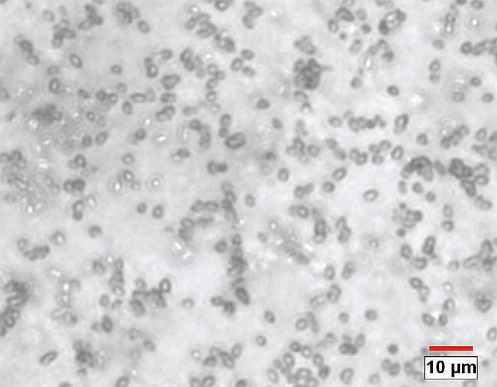
Abbildung 1. Originalbild mit 400-facher Vergrößerung (0,1176 Micron/Pixel). Dieses Bild zeigt ein typisches Feld, bei dem viele Partikel außerhalb des Fokus liegen.
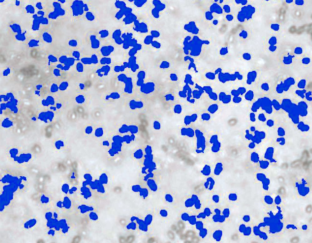
Abbildung 2. Bei einem ersten Durchlauf wurden die im Fokus liegenden Partikel ermittelt. Artefakte wurden eliminiert und es wurde eine erste Messung der verbleibenden Partikel und Agglomerationen durchgeführt.
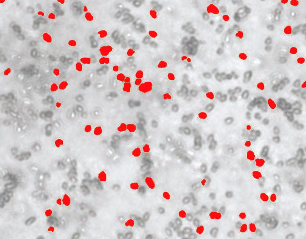
Abbildung 3. Alle Partikel und Cluster wurden nach Rot kopiert und weiter separiert. Nur die Komponenten, die mit großer Wahrscheinlichkeit aus einem Partikel bestanden, wurden beibehalten und es wurde eine zweite Messung durchgeführt.
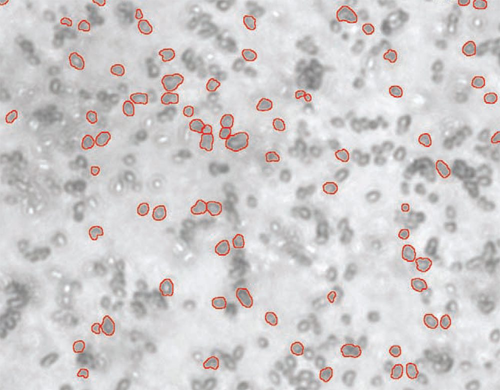
Abbildung 4. Umriss der Partikel, gemessen in der roten Bitebene. Im Umrissmodus lassen sich alle separierten Partikel erkennen.
ZWECK
Zeigt, dass das Clemex Vision Bildanalysesystem eine Partikelgrößen- und -formmessung bei Augentropfenproben mit sehr feinen Partikeln (~1 bis 5 µm) durchführen kann. Partikel dieser Größe bringen die optische Mikroskopie an ihre Grenzen. Das erklärt die Schwierigkeiten, bei dieser Probe scharfe Bilder zu erhalten.
ERGEBNISSE
Für jede Komponente wurden Länge, Kreisdurchmesser, Breite und Ellipsoidvolumen gemessen. Während des Durchlaufs wurden automatisch Statistiken und Diagramme generiert und für die gesamte Probe zusammengefasst. Mit seinen vielseitigen und leistungsstarken Funktionen konnte Clemex Vision die erforderlichen Informationen ermitteln und die in den Augentropfen enthaltenen Partikel quantifizieren.


