
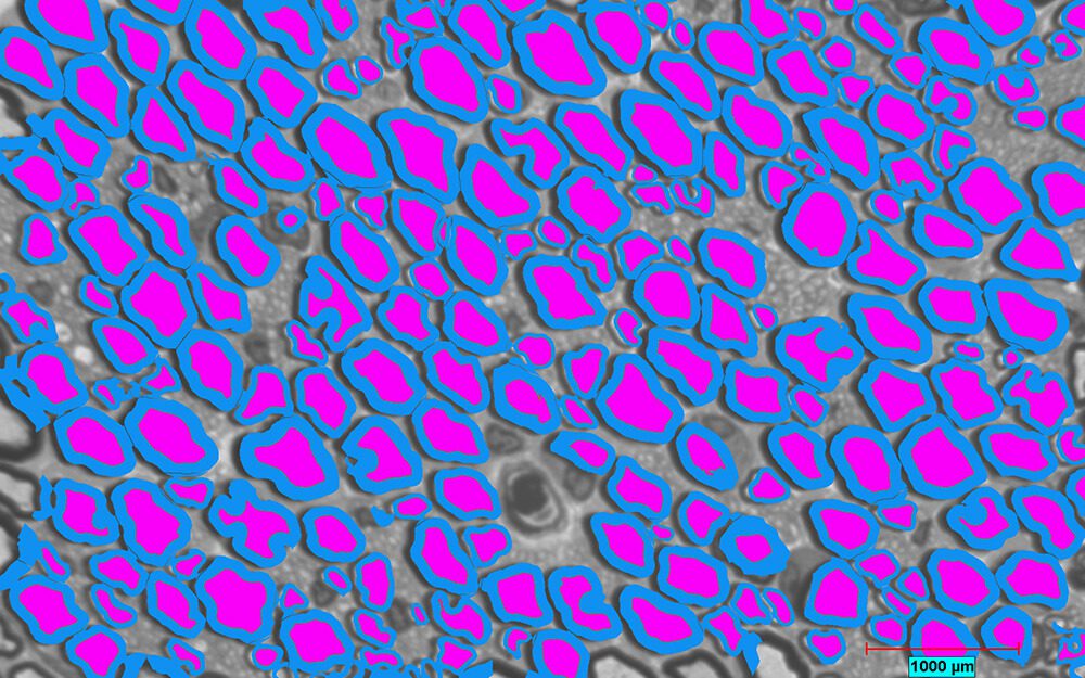
Fig. 1 Nerve profiles detected in blue (myelin) and magenta (axon) using Clemex Vision PE software.
The role of Clemex software in this study
Clemex Vision PE was used to detect nerves separately from background non nerve profiles. The software was able to measure the area of each component – myelin (blue) and axon (magenta). Combining the two colors into another binary plane, the software could also measure the total fiber width and area of axon + myelin. Adding other measurement parameters, such as circular diameter, aspect ratio, or roughness is easily done within the software.
Authors: Daniel A Hunter, Deng Pan, Matthew D Wood, Alison K. Snyder-Warwick, Amy M. Moore, Eva L Feldman, Susan E Mackinnon, Michael J Brenner
Abstract
Background: Stereology and histomorphometry are widely used by investigators to quantify nerve characteristics in normal and pathological states, including nerve injury and regeneration. While these methods of analysis are complementary, no study to date has systematically compared both approaches in peripheral nerve. This study investigated the reliability of design-based stereology versus semi-automated binary imaging histomorphometry for assessing healthy peripheral nerve characteristics.
New Method: Stereological analysis was compared to histomorphometry with binary image analysis on uninjured sciatic nerves to determine nerve fiber number, nerve area, neural density, and fiber distribution.
Results: Sciatic nerves were harvested from 6 male Lewis rats, aged 8–12 weeks for comprehensive analysis of 6 nerve specimens. From each animal, sciatic nerve specimens were fixed, stained, and sectioned for analysis by light and electron microscopy. Both histomorphometry and stereological peripheral nerve analyses were performed on all specimens by two blinded and independent investigators who quantified nerve fiber count, fiber width, density, and related distribution parameters.
Comparison with existing methods: Histomorphometry and stereological analysis provided similar outcomes in nerve fiber number and total nerve area. However, the light microscopy, but not electron microscopy, stereological analysis yielded higher nerve fiber area compared to histomorphometry or manual measurement.
Conclusion: Both methods measure similar fiber number and overall nerve fiber area; however, stereology with light microscopy quantified higher fiber area. Histomorphometry optimizes throughput and comprehensive analysis but requires user thresholding.
Request full article from ResearchGate