Sphere characterization
One Scanning Electron Microscope-Secondary Electron (SEM-SE) image of spheres in BMP format is submitted for analysis.
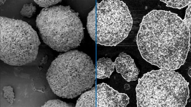
Figure 1. Left: Original image. Right: The original image is modified by a gray transformation to outline the spheres.
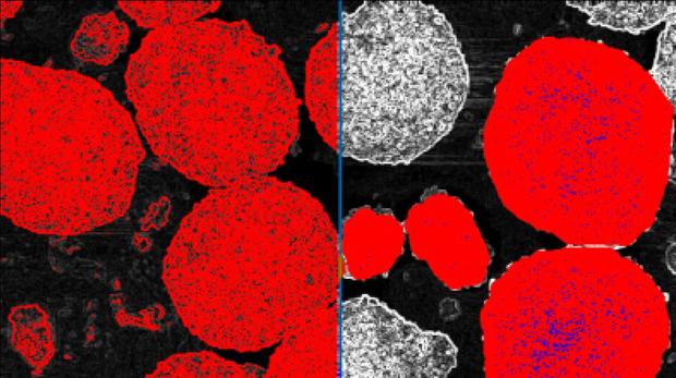
Figure 2. Left: The sphere outlines are binarized into red bitplane by Gray Thresholding. Right: Final detection of spheres (red) and pores (blue) overlay against the modified gray image.
Clemex solution for sphere characterization
Solution
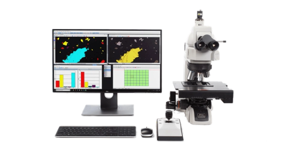
Demonstrate the ability of the Clemex Vision image analysis system to discriminate and measure the spheres and their pores. The methods and operations used are discussed in the report linked at the bottom of this page (click the Download PDF link).
Results
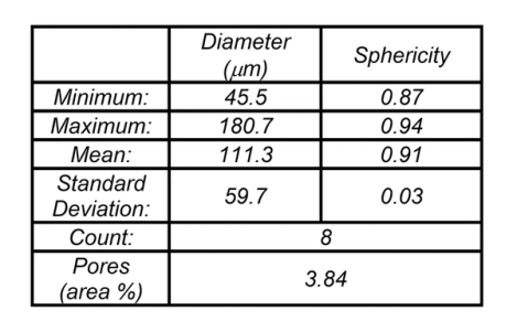
The circular diameter and sphericity of complete spheres as well as the area of pores are measured during the analysis. Final results can be printed directly from Clemex Vision. Raw data are linked to their respective objects for validation purpose. Raw data can also be exported in Excel format.
Let’s discuss your needs, request a quote from our team.
Go back to Application Catalog